Spine Surgery And Back Pain

General Information
Millions of people across India, be an adolescent or an elderly experience back pain. Back pain is listed as the 3rd most common reason to visit a doctor after skin disorders and joint disorders. There are several reasons for a back pain.
Dr. Rohit lists some common reasons as follows-
- Osteoporosis
- Spinal deformities
- Vertebral fracture (s)
- Herniated Disc
- Compressed nerves
- Muscle injury
- Tumors
- Spinal Infections
- Spondylolisthesis
and more….
Back pain can be categorized as acute back pain, chronic back pain and lumbar radicular pain. Generally, acute back pain experienced by the adults lasts for a few days or week and reduces or resolves within this time frame. If the pain lasts longer than 2 months then it is categorized as chronic back pain. Dr. Nalavade aims at treating most of the back problems through non-surgical methods- physical therapy, medication etc.
However, under certain circumstances surgery becomes mandatory. Dr. Rohit Nalavade suggests surgery when-
- Patient experiences persistent and disabling back pain
- Pain radiates to the legs or patient experiences sudden leg weakness or numbness.
- Patient notices absence of reflex responses
- Pain is accompanied with a fever
- Patient is unable to hold their bladder or bowel
The back is divided into 3 regions as shown in the figure-
There are different types spinal surgeries that Dr. Nalavade performs. Some of them are listed below.
The 3 most common type of spine surgeries are-

1) Laminectomy
It is a type of spinal decompression surgery. Spinal decompression can be performed in any region of the back (Cervical spine, Thoracic spine and Lumbar spine). Spinal canal alias Vertebral canal contains a lining through which the nerves pass. Dr. Nalavade explains, due to arthritis, thickened ligaments, bone spurs, enlargement of joints, ruptured or bulged discs, spinal canal becomes narrow as they take up the space through which the which the nerves pass. This narrowing is known as spinal stenosis which causes pain, numbness or weakness of muscles in arms and legs. Hence, Laminectomy is performed by removing lamina (it is the part of the vertebral arch that forms the roof of spinal canal) and bone spurs that enlarges the spinal canal and relieves pain.
Laminectomy is a 6 step procedure, carried out as follows –
– Incision
Incision is made in the back precisely over the affected vertebrae. The length of the incision depends on the number of laminectomies that are going to be performed. However, minimally invasive procedures generally use smaller incisions compared to the open surgery. Dr. Nalavde then spilts the back muscles and move it to the either sides to gain access to the lamina of each vertebra.
– Laminectomy
In this step, lamina is removed using either a drill or bone biting tools. Then thickened ligament flavum – a short ligament that connects the laminae of adjacent vertebrae- is removed. This procedure is repeated for every affected vertebrae.
– Decompressing the spinal cord
Removal of lamina and ligamentum flavum makes the protective covering of spinal cord visible. The protective sac of the spinal cord and spinal nerves are drawn back to eliminate the bone spurs.
– Decompressing the spinal nerve
There are small joints between and behind the each vertebrae which are known as facet joints. These joints are present in the pair of two on each level of vertebral column. Since these, facet joints are directly over the nerve roots, they’re trimmed to give more space to the nerve roots.
– Performance of other surgeries
There are small joints between and behind the each vertebrae which are known as facet joints. These joints are present in the pair of two on each level of vertebral column. Since these, facet joints are directly over the nerve roots, they’re trimmed to give more space to the nerve roots.
– Closure
Sutures or staples are used to sew the muscle and skin incisions.
2) Spinal Fusion
Spinal Fusion is a procedure that involves fusing two or more vertebrae in the spine. It is usually done to correct spine deformities (scoliosis), reduce bank pain that causes weakness in the spine, stabilize the back after the removal of damaged disc (Herniated disc) or to stabilize the back after the spinal fracture. It can be performed in any region of the spine (Cervical spine, Thoracic spine and Lumbar spine). Sometimes, when the pain radiates to legs or arms, Laminectomy (decompression) is performed along with Spinal Fusion.
The procedure of Spinal Fusion involves 3 steps as follows-
– Incision
To gain access to the vertebrae that is being fused, Dr. Nalavade makes incision. Vertebrae can be accessed through three different approaches known as Anterior Approach, Posterior Approach and Lateral Approach. Under Anterior approach, incision is made in lower abdomen (for lumbar fusion) or in front of the neck (for cervical fusion). Since, spine is approached from the front it is known as Anterior Approach. In Posterior Approach, spine approached from the back and incision is made in middle of lower back or in the neck. Under Lateral Approach, the spine is approached from the side, incision is made on either side of the spine. The right approach for any surgery depends on the nature and location of the disease.
– Bone Grafting
Small pieces of bones are arranged between the spaces of the vertebrae to be fused. The small piece of bone material is known as bone graft. Bone graft can be acquired from the bone bank or patient’s pelvis. If you’re undergoing decompression procedure with spinal fusion, Dr. Nalavde may use the bone from where it is compressing nerves to the area where vertebrae needs to be fused, this type of graft is known as local autograft. Nowadays, different types artificial bone materials are used to carry out spinal fusion that aids bone growth and speeds fusion. Dr. Rohit Nalavade suggests the type of bone graft material for your surgery depending on your condition.
– Fusion
The final step is fusion. To permanently fuse the vertebrae together, bone graft is used between the vertebrae. In most of the cases rods, plates and screws are used to hold the spine still.
3) Discectomy
Dr. Rohit Nalavde explains that Spinal column is made of individual bones that are named as vertebrae. These vertebrae are placed on top on each other. In between them they have shock absorbing discs. However, these discs may bulge out or become herniated, which can cause pain in legs, arms or buttocks or nerve weakness that would lead to trouble standing or walking. Therefore, Discectomy is performed by removal of proportions of the disc that irritate or press nearby nerves.
Different types of discectomy are:
- Microdiscectomy
- Percutaneous Discectomy
- Endoscopic Discectomy
- Laser Discectomy
The group or collection of nerves at the end of the spinal cord, resembling a horse tail, is called as Cauda Equina. Under Cauda Equina Syndrome, roots of the nerve bundle get squeezed at the end of spinal cord. It is caused by herniated discs which may lead to pain and weakness in the legs, loss of bowel or bladder control etc.
The procedure involves the following steps-
- The incision is made precisely over the region where the disc is damaged. Dr. Nalavade gains access to the affected region by splitting the back muscles using dilators or retractors.
- In the next step, laminectomy is performed, the lamina is removed which forms a window through which spinal nerves can be viewed.
- Then the herniated disc is removed along with other fragments of the disc that may have been displaced or likely to get displaced.
- Finally, Sutures or staples are used to sew the muscle and skin incisions.
Other types of spine surgeries are-
4) Anterior Cervical Discectomy and Fusion
Anterior Cervical Discetomy and Fusion is a type of neck surgery. The surgery is performed to remove the damaged disc, Dr. Nalavde reaches the disc through the throat area from the front. By retracting the neck muscles and moving the muscles, trachea and esophagus on the either side; he gains access to the disc and vertebrae. After the removal disc, space between the vertebrae is cleared. To prevent the vertebrae from rubbing against each other or collapsing, Dr. Nalavade excutes spinal fusion by placing the bone graft in the emptied space. The bone graft and vertebrae are fixed using plates and screws.
ACDF is carried out when the discs bulge out or becomes herniated, bone spurs cause pain or ligaments inflame causing canal stenosis.
5) Kyphoplasty
If you have osteoporosis, a compression fracture is something you might experience. Spinal compression fractures occur usually in the thoraric region of the spine. However, it may occur in lumbar region as well. Kyphoplasty is performed to treat spinal compression fractures. The aim of kyphoplasty is to bring stability of vertebra, reduce pain that arises from fracture and restores the vertebrae back to the normal height.
The procedure involves making an incision precisely on the affected region. Through the counsel of X-ray, Dr.Nalavade proceeds to insert a narrow tube into one side of the fractured vertebra. Then through the tube Dr. Rohit, inserts a balloon into the vertebrae and slowly inflates it. Fracture is alleviated as the balloons get inflated, causing the displaced bones to return back to their original position. It further compresses the soft inner bone to create cavity in the vertebrae. After that, balloon is removed, PMMA, a type of bone cement, is used to fill the cavity. The cement dries and hardens quickly, stabilizing the bone. In other types of kyphoplasty, instead of a balloon a net or ball maybe used to create the cavity.
6) Vertebroplasty
People with osteoporosis or cancer are familiar with compression fractures. Spinal compression fractures usually occur in the thoraric region of the spine. However, it may occur in lumbar region as well. Vertebroplasty is performed to treat spinal compression fractures. The aim of Vertebroplasty is to bring stability of vertebra, reduce pain that arises from fracture and strengthen the vertebra.
During the surgery, the skin above the injected region is punctured using a biopsy needle. With the help of X-ray, the biopsy needle is inserted deep in the vertebra using rotator motion or a tapping mallet. PMMA, a type of bone cement, is injected in the fractured vertebra. The cement fills the cracks in the bone, needle is removed and cement quickly hardens thereby stabilizing the fractured bone. Puncture in the skin is enclosed with a bandage.
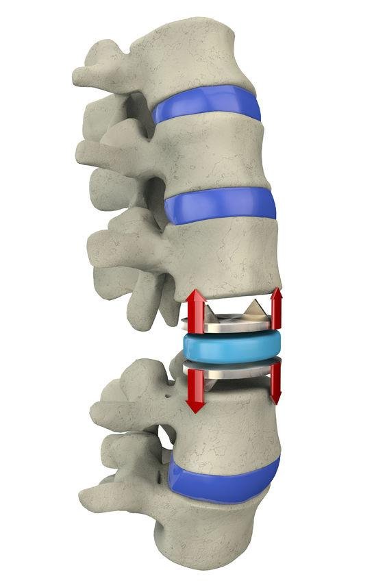
7) Artificial Disc Replacement
Spinal Fusion has been the conventional surgery to provide stability to vertebrae after execution of discectomy surgery. Artificial Disc Replacement is the new type of surgery that replaces Spinal Fusion. Instead of fusing the bone graft with the vertebrae, artificial discs are used in the emptied space. The artificial discs are made of metal and plastic materials. It is a procedure that utilizes anterior approach. Artificial discs allow motion whereas the fusion restricts motion. However, ADR is not recommended to you if have multiple level degenerative disc disease or neurological compression. It is best for young people with non-significant arthritis.

I offer surgical treatments known as minimally invasive surgery. Under MIS procedure, smaller incisions are made and it ensures quicker recovery.
Book a consulting with me now!!!
Our Services
Menu